What is the anatomy of the knee?
The human knee is complex hinge-joint which allows individuals to walk, run, squat, sit and jump. The anatomy of the knee allows the joint to handle large amounts of demand and stress each day. The flexibility of this joint can, however, make the knee vulnerable to injury. Dr. Armando Vidal, orthopedic knee specialist, is an expert at diagnosing and treating knee injuries and pain. Patients in Vail, Aspen and surrounding Denver, Colorado communities can depend on Dr. Vidal to help them return to their busy and active lifestyle.

What makes up the knee joint?
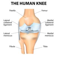
The anatomy of the knee is made up of bones, ligaments, tendons, cartilage, muscles and bursa.
Bones: Three bones meet to form the knee joint, they are:
- Femur (thigh bone)
- Tibia (shin bone)
- Patella (knee cap.)
Cartilage: Two types of cartilage make up the knee joint:
- Meniscus – or Menisci – Two C-shaped discs which act as “shock absorbers” in the knee, stabilizing the joint between the femur and the tibia so the knee can bend and flex without the bones rubbing together. The menisci are rough, rubbery and contain nerves that help improve balance and stability by distributing weight evenly. Meniscus deficiency can lead to wear of the articular cartilage and instability in certain cases.
- Medial Meniscus – larger of the two menisci in the knee, found on the inside of the knee joint.
- Lateral Meniscus – smaller cartilage, found on the outside of the knee joint.
- Articular cartilage – found on the ends of the femur, tibia and the back of the patella. Often described as the white, shiny substance on the end of a chicken bone, this slippery layer acts as a shock absorber and helps the knee bones glide smoothly across each other as the knee is bent or straightened. This tissue is highly complex and one of the most specialized tissues in the body.
Ligaments: Ligaments hold bone to bone, acting like strong ropes in the knee, providing strength and stability:
- Anterior Cruciate Ligament (ACL) – forms ½ of an “X” with the PCL ligament, traveling from the front of the tibia to the back of the femur.
- Posterior Cruciate Ligament (PCL) forms the other ½ of an “X” with the ACL, travels from the back of the tibia to the front of the femur.
- Medial Collateral Ligament (MCL) travels down the inside of the knee.
- Lateral Collateral Ligament (LCL) travels down the outside of the knee.
Tendons: Muscles are connected to bones by tendons. These bands of soft tissue are similar to ligaments, but instead of connecting bone to bone, they connect muscle to bone. Knee tendons include:
- Patellar tendon – attaches the tibia to the patella (shin bone to the kneecap)
- Quadriceps tendon – connects the muscles in the front of the thigh to the patella. Largest tendon in the knee joint.
Muscles: While the quadriceps muscles and the hamstrings are not technically part of the actual knee joint, they do play an important roll in giving the knee flexibility and strength.
- Quadriceps – Four muscles in the front of the leg that help straighten the knee.
- Hamstrings – Three muscles in the back of the knee that help bend the knee.
- Gluteal muscles – Gluteus maximus and gluteus minimus, also known as the glutes or the buttocks are important for the knee’s positioning. Also called hip abductor muscles, are important in the function of the joint behind the knee cap known as the patellofemoral joint.
Bursae: The knee joint contains approximately 14 bursae. Bursae are small, fluid-filled sacs that allow tendons to move fluidly over bony prominences. Think of how different the skin on the front of your knee is from the skin in other parts of the body. It is the bursa between the knee cap and that skin that allows it to move so freely.
What are common knee injuries?
The most common knee injuries for patients in in Vail, Aspen and the surrounding Denver, Colorado communities are:
There are many approaches to treating knee pain and injury. Often, common knee injuries can be treated by a non-surgical approach. If surgery is needed, Dr. Vidal and his sports-medicine team will determine the proper surgical technique to alleviate pain, improve knee stability and return the anatomy of the knee to its proper condition.
Locations
180 S Frontage Rd W
Vail, CO 81657
226 Lusher Court
Ste 101
Frisco, CO 80443
322 Beard Creek Road
Edwards, CO 81632


