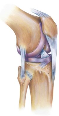
What is the posterolateral corner of the knee?
Not too long ago, the posterolateral corner of the knee, also called PLC, was referred to as the dark side of the knee. This was because of the limited understanding of the biomechanics, structures and treatment options for this area of the knee. In recent years, orthopedic surgeons, like Dr. Armando Vidal, have completed a number of studies which have led to a greater understanding of the PLC and associated reconstruction techniques. Dr. Vidal and his colleges at The Steadman Clinic, serving Vail, Aspen and the surrounding Denver, Colorado communities are thought leaders and experts in treating the posterolateral corner.

What makes up the PLC?
Think of the posterolateral corner as a miniature bit of armor, guarding against lateral or external forces. Located on the outside of the knee joint, its job is to stabilize the knee and protect the joint from buckling laterally. Three main static stabilizers make up the PLC, those are; the fibular collateral ligament (FCL) – sometimes called the lateral collateral ligament (LCL), the popliteus tendon (PLT) and the popliteofibular ligament (PFL). These stabilizers are attached to the bones that make up the “corner”; the tibia, fibula and the femur. (Shin bone, smaller lower-leg bone and long thigh bone, respectively.)
How does the posterolateral corner become injured?
A posterolateral corner injury rarely happens in isolation; only 28% of all PLC injuries involve just the structures in the posterolateral corner. An injury of this severity often includes damage to other ligaments such as the PCL (posterior cruciate ligament) and/or ACL (anterior cruciate ligament). A posterolateral corner injury most often occurs with athletic trauma, severe falls and car accidents. Contact sports like football or hockey or other activities where the knee can experience severe trauma are often the main contributors to a PCL injury.
What are the symptoms of a posterolateral corner injury?
Symptoms often reported when there is damage to the PLC are:
- Swelling of the knee located on the lower, outside area (posterolateral), can be minimal.
- Pain on along the outside of the knee
- Knee instability from side-to-side
- Difficulty when pivoting, turning or twisting
How is a PLC injury diagnosed?
Dr. Vidal will obtain a complete patient medical history, including events that lead to the knee injury. He can perform several physical exam tests to determine the stability of the knee and if there is an injury present. These tests may include:
- Dial Test – Turning the foot outward to determine the rotation of the knee. Excessive rotation indicates a PLC injury.
- Posterolateral Drawer Test – Dr. Vidal will bend the hip and knee to check along the outside for tibia movement, or if it slides to the side while bending.
- Varus Stress Test – Determines the amount of increased rotational instability and the true amount of lateral compartment gapping
- Reverse Pivot Shift: Another test where Dr. Vidal bends the knee while applying slight pressure and rotation. Since some rotation is normal, he will compare the decree of shift with the healthy knee.
Imaging can be tricky in PLC injuries. In acute injuries, an MRI can be reliable in indicating that an injury occurred, but in and of itself does not tell you the severity of the injury or if these structures are competent. In chronic injuries or in the setting of revision surgery – an MRI is not an accurate measure of how competent the PLC really is. In these cases, a radiographic stress test will be performed. This essentially means that Dr. Vidal or one of his trained assistants will stress your knee (and your normal knee as a comparison) similar to what was done on physical examination and obtain an X-ray while performing the stress. Any asymmetry will be measured to determine functional integrity of the PLC. This is a key test and is the only objective measure of PLC integrity.
How is a posterolateral corner injury treated?
Minor PLC can heal without significant impact. Complete PLC injuries require surgery to reconstruct the soft tissue damage. This surgery is typically done as an open procedure to reconstruct the knee joint and can be done in conjunction with other arthroscopic procedures such as ACL and /or PCL reconstruction.
Complex Knee Specialist
Damage to the PLC is a serious injury resulting in instability and pain in the knee. Over the years, experts such as Doctor Armando Vidal have studied the PLC, allowing for more advanced, more successful treatment options to be practiced. Patients in Vail, Aspen, and the surrounding Denver, Colorado communities who have experienced a PLC injury should see Dr. Vidal for diagnosis and a specialized treatment plan that will ensure the best outcome for the patient’s particular injury. Contact Dr. Vidal’s team today!

Locations
180 S Frontage Rd W
Vail, CO 81657
226 Lusher Court
Ste 101
Frisco, CO 80443
322 Beard Creek Road
Edwards, CO 81632


